Blue Star STING Quick Learn


We would like to be as silent as possible here!
Pictures are going to be your guide and just here and there we will give some textual instructions.
One picture is worth 1000 words. And we hope that pictures are descriptive;
Enjoy the cruise! And, by the way, if you wish to be faster in Quick_Learning, try DIMOND STING WARP Speed Results! See also this application! And see how to obtain images as in our publication published by NAR Special Web server issue (2003).
We highly recommend that the user gets acquainted with information that PDB files are carrying; use PDB_Metrics for this purpose!


Basic commands Modules
Before you start
Initialize STING Millennium
Action example
Activate menu options
Search Pattern and Search Neighbors
Build Surface and Interface
Ramachandran plot   ConSSeq   STING_TGZ
Hornet   Java Ca-Ca/Cb-Cb Distance Plot   MSSP 2D plot
Scorpion   XY Graphics Java Plot   STING_DB QA
Formiga   Sting Report   JPIV
Graphical Contacts   Java Protein Dossier   AA Co-evolution
IFR Contacts   Java Table of Contacts   Protein-ligand Contacts
STINGpaint   Phylogenetic Tree   Contact Distance Map
PDB_Cartoon   STING MSA   Protein Contacts Difference
Protein Dossier   DNA Contacts   TopSiMap
Topologs 100   Topologs ASTRAL 40   STING Enzyme Classification


STING Millennium Modes Data Parsing --- Blue Star STING: Links
Multiple Sequence Alignment with single 3D structure
Multiple Structure Alignment with sequences
LINKING to STING and its components:
Direct your call to Servers in Brazil
Direct your call to Servers in USA
PDB_Metrics   Secondary Structure
Structure Information   Enzyme Information
Structure Classification   Sequence neighbours
Structure Genomics   Surface/Structure
Cavity Report   Surface Validation
Domains based on Structure   User input required: Links

STING Millennium Quick LearnBasic commands

Before you start

To make right choice of your browser, please see here!

To see the 3D structure you need to download and install the CHIME plugin

Also, chances are very small, but you may be interested in one of "very specific PDB files" and there will be very specific STING response! There are also "very creative residue numbering in PDB files". See about known STING bugs here! Also, get acquainted with general characteristics of PDB through our PDB_Metrics!

Let us analyze 1dhk.pdb (alpha amylase in complex with inhibitor):


Initialize STING Millennium

Left Image:
There are two possible ways to initialize STING Millennium in basic mode (other two modes are described in STING Millennium Quick Learn Modes):
A) Browsing at your local computer for the "pdb" format file containing your molecule of interest, and
B) Indicating 4 letter code of the "pdb" format file {in this case 1dhk.pdb was loaded} .
In this latter case, pdb file is loaded from the location where STING Millennium Server is installed;
Option B was used for this Quick Learn session.

Right Image:
This is how STING Millennium will respond!

Please learn more about STING Millennium basic features!

Action example


We hope that no words are necessary here! To get from the display at the upper right image to the lower left image, just follow magenta line instructions!

Left Image:

Ribbons !
Note: water molecule positions are indicated by red wireframe presentation.
Note #2: Try here experimenting and see what type of img you will get by successively clicking on the REFRESH button!

Right Image:
Ligand on!

See more of ACTION menu options!


Activate menu options:

  • It is important to know that the menu option "Int Surface" and options in "Modules" menu: "Formiga" and "IFR Contacts", will be ACTIVATED only if the user first does definition of the Interface by executing "Interface chain" option available in the STING Millennium "Windows" menu option.
  •  


    Left Image:
    This is the difference between inactive and active menu option:
    -on the left is the inactive menu option,
    -on the right is the same menu option, but now activated!
    Right Image:
    To activate inactive menu options, the user should follow magenta line instructions!.

    See more options on Interface On menu!
    See more options on Int Surface menu!


    Search Pattern and Search Neighbors


    Left Image:
    Search Pattern: simple and effective!
    Right Image:
    Search Neighbors:
    To define neighbors, one would first need to define the central amino acid. This is done by marking (red CPK) of Gly (in this case). Afterwards, the user defines the radius of the sphere where either "Alpha"-carbon to "Alpha"-carbon distances are measured, or Last Heavy Atom in side chain to LHA distances are measured. Neighbors are graphically presented in the chosen color (orange, in this case) after the user clicks on the same central amino acid, with the right or left mouse button.


    Build Surface and Interface

    Left Image:
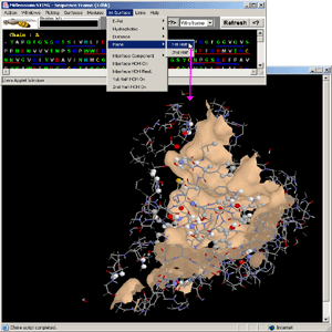
    At the lower left inset, we choose to display gold color "Plane" surface. The other two options are: EP (stands for Electrostatic Potential), and Hyd (stands for Hydrophobicity). In those two cases, the surface would be painted with color coded scales representing for each amino acid the corresponding feature: EP and Hyd, respectively.


    Image bellow:

    In this example, the user can observe just the "1st half" of the interface as a solid surface. This is the part of the interface designated at "Activate menu Options - Right Image" as chain A. The spacefill presentation is done only for the atoms that are within contact distance from the atoms belonging to the Interface Forming Residues (IFR) from the opposite side of the presented surface. All IFRs are displayed in wireframe presentation.
    Go to Quick Learn: Modules
    Go to Quick Learn: Modes